Chlorophyll-a synthase in two non-photosynthetic Cyanobacteria of the Sericytochromatia

One of the most exciting and intriguing findings in the subject of the evolution of oxygenic photosynthesis in the past few years is the discovery of the non-photosynthetic early-branching Cyanobacteria: the Vampirovibrionia (formerly Melainabacteria) (Soo et al. 2019) and the Sericytochromatia. They are considered to be within the phylum Cyanobacteria. The other really interesting new finding is the Margulisbacteria, which is considered to be the sister phylum to Cyanobacteria.

All the genomes of the known representatives of these novel clades are non-photosynthetic. Many of them live in environments where light cannot reach or live as symbionts. One of the very spectacular bugs in these new clades, for example, is Termititenax. This is a margulisbacterium that attaches to an ectosymbiotic spirochete of an oxymonad protist in the gut of termites (Utami et al. 2019). That is a three-level symbiosis involving four partners! Another interesting strain is Vampirovibrio chlorellavorus (Soo et al. 2015), which is a predatory bacterium that eats the eukaryote Chlorella!

It has been hypothesized that because these new clades do not have any photosynthetic representatives, the photosynthetic Cyanobacteria could have evolved photosynthesis at a relatively late stage, after the divergence of Vampirovibrionia.

I, and others, have said that the lineage from which the photosynthetic Cyanobacteria emerged is directly linked, through an unbroken line of descent, to the origin of photosynthesis itself. I have also said, based on the molecular evolution of the photosynthetic reaction centres, that the earliest stages in the evolution of photosynthesis predate the diversification of the major groups of bacteria. In other words, Margulisbacteria, Sericytochromatia, and Vampirovibrionia, as well as most, if not all bacteria, could have originated from photosynthetic ancestors. In a way very similar to methanogenesis in Archaea, which is thought to be the ancestral trait of the domain and was later lost repeatedly in many clades.

How do we prove however that an organism that today is the symbiont of a spirochete, which is the symbiont of an oxymonad, which is a symbiont of a termite, originated from a photosynthetic ancestor?

If the loss of a process, say a metabolic pathway, was relatively recent. Then, there may be traces of the lost pathway in the genome. Perhaps one of the enzymes in this lost pathway catalysed a type of reaction that could be used for something else and was therefore retained. Loss of photosynthesis in eukaryotes are common at most taxonomic levels, but it is much harder to prove it for bacteria. For example, the algae symbiont, Athelocyanobacterium thalassa, lost 75% of its genome in less than 100 million years (Cornejo-Castillo et al. 2016). Thus, very quickly, any trace of a photosynthetic past may have been erased after the ancestor of these clades became heterotrophic.

There are quite a few proteins in photosynthesis that can catalyse reactions that can be useful outside photosynthesis. So, there is a possibility that some of the free-living non-photosynthetic Cyanobacteria that retain larger genomes, from those lineages that did not become symbionts and/or experienced strong genome-size reductions, still contain remnants of photosynthesis proteins that have been co-opted for new functions.

Here I report two sequences that branch within the known diversity of bacteriochlorophyll a and chlorophyll a synthases, known as BchG and ChlG respective, in the metagenome-assembled genomes (MAGs) of two strains of Sericytochromatia. BchG and ChlG catalyse the final step in the synthesis of chlorophylls and bacteriochlorophylls and have the job of attaching the characteristic tails of these pigments to the tetrapyrrole ring.

I found one sequence using AnnoTree. There are four Sericytochromatia genomes and 53 Vampirovibronia genomes available there. I searched using the KEGG code K04040.

This one is found in the MAG GCA_002083825.1, contig DAZV01000039.1_72, which encodes 79 genes and it was published in Soo et al. (2017). The metagenome came from a coal bed methane well.

The second sequence I found using BLAST on the refseq database. It was the top hit when using the above sequence as query. This MAG was published in Parks et al. (2018) and the metagenome is from sediments. This is uncultivated Cyanobacteria bacterium UBA8530.  This is the sequence and it was found in a contig encoding 12 genes.

I did not find BchG or ChlG in the Margulisbacteria MAGs available in AnnoTree.

The evolution of these synthases is very interesting because the enzyme is related to a key enzyme in Archaea, digeranylgeranylglyceryl phosphate synthase (DGGGPS), that is central to the discussions on the nature of the LUCA. I wrote about this a bit in a previous post: https://www.tanaicardona.com/blog/chlorophylls-the-origin-of-membranes-and-bioenergetics

These synthases are also related to other class of enzymes: for example, UbiA used in the synthesis of quinones or CyoE used in the synthesis of some types of heme.

The BLAST of these two sequences only retrieved (bacterio)chlorophyll synthases from phototrophs and suggested a bit of a phylogenetic distance. I collected some sequences and ran a Maximum Likelihood tree with PhyML.

Phylogeny of ChlG and BchG.

Phylogeny of ChlG and BchG.

We can see in the figure above that the sequences from these strains branched together as sister to that from Heliobacteria. Although that position has no statistical support (0.1). Overall the level of sequence identity between the different BchG/ChlG sequences is in the region of just under 30%, but they align very well. At a sequence level they can be easily distinguished from other enzymes in the extended UbiA family due to the very large distances between the types.

Some Chlorobi and Chloroflexi have two BchG.

We can see that the sericytochromatian branches are the longest, more than twice as long as the standard synthases, suggesting faster rates of evolution.

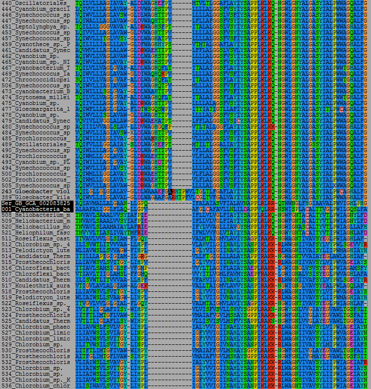
Unfortunately, we do not know a lot about the mechanism of function of these enzymes. There are no crystal structures and I was not able to find how chlorophylls bind or anything of the sort. Looking at the sequences, the sericytochromatian ones share a peculiar 13 amino acid gap with the BchG of the anoxygenic phototrophs, compared to a unique 13 amino acid insertion relative to the Gloeobacter sequences, and a 7 amino acid insertion compared to all other cyanobacterial sequences.

An alignment showing some interesting indels. This is just a part of the alignment. The sericytochromatian sequences are highlighted in black.

An alignment showing some interesting indels. This is just a part of the alignment. The sericytochromatian sequences are highlighted in black.

There is however no biochemical or structural evidence that could reveal what these differences mean in term of function. Overall, it appears as if they are more closely related to BchG than ChlG.

The large branches would be consistent with these sequences acquiring a novel function.

These synthases are membrane proteins, so I had a look at the secondary structure prediction and it turned out that overall the sericytochromatian sequences seem to be more like the cyanobacterial and chloroflexi sequences in topology, than those in other anoxygenic phototrophs, sharing 8 well-defined transmembrane helices.

transmembranes.jpg

Interestingly, unlike other UbiA-like proteins DGGGPS also has 8 predicted helices, which may therefore represent the ancestral state.

In any case, these are just superficial similarities and differences.

I had a look at the other genes encoded in the contigs and there was nothing that would stand out (to me) that could give some insight on the possible role of these proteins in the Sericytochromatia. In GCA_002083825.1, the sequence was two or three genes downstream of HemB, which is an enzyme in the synthesis pathway of porphyrins, but HemB was not in the UBA8530 contig. No other enzymes of the porphyrin synthesis pathway were found in the contigs.

Microbial mats from lake Vanda are known to contain a substantial number of free-living Vampirovibronia and Sericytochromatia strains. So, I BLASTed the sequence from GCA_002083825.1 against the Vanda metagenomes in the IMG/MER database, but I did not find any sequence that matched the Sericytochromatia ones, at least not within the complete sequences. I did get hits for Cyanobacteria and some of the other phototrophs as well as a eukaryotic alga.

It cannot be told whether these two sequences are ancestrally sericytochromatian or not, but these two strains, as well as their unique sequences, appear to be quite distant apart, so it may be an ancient legacy of a phototrophic past. They might just as well have been picked up from DNA of true phototrophs at some early point in time before they diverged and went on their separate pathways, as it is the case for genes encoding protochlorophyllide reductase found in two genomes of subsurface Altiarchaea: http://tanaiscience.blogspot.com/2017/05/a-new-undiscribed-clade-of-phototrophic.html

Given that there are very few Sericytochromatian MAGs available, only 9 in the Genome Taxonomy Database; and given that two of these had (B)ChlG, then it may be that this protein is somewhat widely distributed within this clade.

An apparently early-branching BchG was actually found in a contig from an archaeon and it was shown to have chlorophyll synthase activity (Meng et al. 2009). It would be interesting to do the same experiment with the sericytochromatian sequences! I might repeat the tree above at a later stage including this archaeal sequence, after doing a more extensive search of the metagenomes, and as I become more familiar with these type of enzymes and their other relatives.

I am compiling a number of candidate proteins that could be informative regarding the loss of photosynthesis in these clades for further study. Stay tuned for more.


References

Cornejo-Castillo, F. M., A. M. Cabello, G. Salazar, P. Sanchez-Baracaldo, G. Lima-Mendez, P. Hingamp, A. Alberti, S. Sunagawa, P. Bork, C. de Vargas, J. Raes, C. Bowler, P. Wincker, J. P. Zehr, J. M. Gasol, R. Massana and S. G. Acinas (2016). "Cyanobacterial symbionts diverged in the late Cretaceous towards lineage-specific nitrogen fixation factories in single-celled phytoplankton." Nat Commun 7. DOI: 10.1038/ncomms11071.

Meng, J., F. Wang, F. Wang, Y. Zheng, X. Peng, H. Zhou and X. Xiao (2009). "An uncultivated crenarchaeota contains functional bacteriochlorophyll a synthase." ISME J 3(1): 106-116. DOI: 10.1038/ismej.2008.85.

Parks, D. H., M. Chuvochina, D. W. Waite, C. Rinke, A. Skarshewski, P. A. Chaumeil and P. Hugenholtz (2018). "A standardized bacterial taxonomy based on genome phylogeny substantially revises the tree of life." Nat Biotechnol 36(10): 996-1004 DOI: 10.1038/nbt.4229.

Soo, R. M., J. Hemp and P. Hugenholtz (2019). "Evolution of photosynthesis and aerobic respiration in the cyanobacteria." Free Radic Biol Med. DOI: 10.1016/j.freeradbiomed.2019.03.029.

Soo, R. M., J. Hemp, D. H. Parks, W. W. Fischer and P. Hugenholtz (2017). "On the origins of oxygenic photosynthesis and aerobic respiration in Cyanobacteria." Science 355(6332): 1436-1440. DOI: 10.1126/science.aal3794.

Soo, R. M., B. J. Woodcroft, D. H. Parks, G. W. Tyson and P. Hugenholtz (2015). "Back from the dead; the curious tale of the predatory cyanobacterium Vampirovibrio chlorellavorus." PeerJ 3: e968. DOI: 10.7717/peerj.968.

Utami, Y. D., H. Kuwahara, K. Igai, T. Murakami, K. Sugaya, T. Morikawa, Y. Nagura, M. Yuki, P. Deevong, T. Inoue, K. Kihara, N. Lo, A. Yamada, M. Ohkuma and Y. Hongoh (2019). "Genome analyses of uncultured TG2/ZB3 bacteria in 'Margulisbacteria' specifically attached to ectosymbiotic spirochetes of protists in the termite gut." ISME J 13(2): 455-467. DOI: 10.1038/s41396-018-0297-4.

 

My two pesos on the metagenome anomaly controversy

On the 9th of August (2019) a team of researchers from German, Chinese and Indian institutions uploaded a rather controversial preprint to the bioRxiv:

Anomalous phylogenetic behavior of ribosomal proteins in metagenome assembled genomes by Garg et al. https://doi.org/10.1101/731091

The team challenged the idea that metagenome-assembled genomes (MAGs) represent real organisms, but suggested that these are “unnatural constructs, genome-like patchworks of genes that have been stitched together into computer files by binning.

Needless to say, it created a little twitter storm.

The challenge focused on two major groups of prokaryotes, the Asgard archaea and the Candidate Phyla Radiation (CPR) of bacteria. These two clades have been mostly erected on metagenome projects, although independent evidence for the existence of these groups have been provided through single-cell genomics (Rinke et al. 2013, full references at the end of the post) or the enrichment of a Loki archaeon culture and the sequencing of its genome (Imachi et al. 2019), for example.

The challenge was grounded on phylogenetic evidence. The authors show that the ribosomal proteins found in MAGs of Asgard archaea and CPR bacteria do not retrieve consistent phylogenies, as if different ribosomal proteins would have been “stitched together” from very different sources.

Fig. 3 Neighbor-Nets reconstructed from concatenated alignments of 23 ribosomal proteins for archaeal reference samples and archaeal MAGs.(a) The Neighbor-Net of a concatenated alignment of 23 ribosomal proteins in the archaeal reference sample ARS3…

Fig. 3 Neighbor-Nets reconstructed from concatenated alignments of 23 ribosomal proteins for archaeal reference samples and archaeal MAGs.

(a) The Neighbor-Net of a concatenated alignment of 23 ribosomal proteins in the archaeal reference sample ARS3001 shows very little conflict throughout, resulting in a tree-like network with 16 well supported splits (indicated with red dots). (b) A Neighbor net drawn from a concatenated alignment of the same 23 ribosomal proteins from Asgard archaeal MAGs results in a network with a star-like structure. The insets magnify the central area of interest to better highlight the difference of signals of the two networks. Taken from Garg et al. (2019).

So what is going on?

I have to say that this does not surprise me at all. I am not a metagenomics or bioinformatics specialist, but I do use some basic tools often and regularly search both standard genome and metagenome databases for protein sequence of my interest. I always, without exception, find assembly errors in standard genomes from supposedly axenic isolates and in metagenomes.

At first it blew my mind, now I just find it a nuisance. I have written about this before, and here I give you some examples:

Trichoplax adhaerens, a weird animal with a piece of Photosystem I

Contamination of genome projects with DNA from other organisms

A cyanobacterium with an anoxygenic Type II reaction center from purple bacteria? (Contamination)

2. Results – First phylogeny of BchC and we have discovered a new phototroph!

But what really blows my mind now is how easily such anomalies could be confused with horizontal gene transfer. I am so used to these type of problems that the work by Garg et al. appears to me only timely. Indeed… I kind of expected something like this would happen at some point.

And yet, I have myself some evidence that the CPR are a real thing! Last year we published a phylogenetic study of FtsH proteins. We collected over 6000 FtsH subunits across all bacteria and eukaryotes, including 247 CPR sequences and definitely, the CPR made a monophyletic group. However, the CPR FtsH clustered within the standard diversity of life and did not show the level of divergence suggested by Hug et al. (2016). In fact, comparing trees, the phylogenetic position of the CPR FtsH is more similar to that reported in the single-cell genomic study by Rinke et al. (2013) than to the metagenomic study by Hug et al. (2016).

Phylogeny of FtsH proteases. CPR in red.

Phylogeny of FtsH proteases. CPR in red.

If the CPR made a distinct lineage to that which encompasses the classical known diversity of bacteria, then the position of CPR FtsH is rather anomalous, even if horizontal gene transfer is invoked.

This year, in collaboration with two other early-career scientists, we reported a phylogenomic study of a novel group of phototrophs belonging to an uncultivated phylum of bacteria, the WPS-2 or the Candidatus Eremiobacterota. This phylum is entirely made of MAGs, but we found clearly divergent photosynthetic genes, that had some distant affinity to the Chloroflexi. The ribosomal protein tree did place Eremiobacterota near the Chloroflexi, which seemed like a consistent story. Although, we also detected what appears to be significant horizontal gene transfer into this novel clade (Ward et al. 2019).

Interestingly enough, in the preprint by Salcher et al. (2019) an imaging technique was used to spot some Asgard archaea, but the imaged cells look nothing like what Imachi et al. (2019) reported for the isolated Loki archaeon. Is that just a coincidence?

My conclusion is that the truth is perhaps in the middle. Metagenomes do contain strong signals of novel diversity and novel clades, but many, if not most MAGs (?), do contain a very substantial contribution from foreign sequences that is underestimated by assembly quality control protocols... and which leads to anomalous distances and phylogenetic posititions. But that is my non-expert opinion!

I do think the challenge by Garg et al. is entirely valid and justified, and I hope it will trigger a critical revision of metagenomic data and of the conclusions that have emerged from metagenome projects. I will look forward to seeing a counter-attack! :)

References 

Di Rienzi, S. C., I. Sharon, K. C. Wrighton, O. Koren, L. A. Hug, B. C. Thomas, J. K. Goodrich, J. T. Bell, T. D. Spector, J. F. Banfield and R. E. Ley (2013). "The human gut and groundwater harbor non-photosynthetic bacteria belonging to a new candidate phylum sibling to Cyanobacteria." Elife 2. DOI: 10.7554/eLife.01102.

Hug, L. A., B. J. Baker, K. Anantharaman, C. T. Brown, A. J. Probst, C. J. Castelle, C. N. Butterfield, A. W. Hernsdorf, Y. Amano, K. Ise, Y. Suzuki, N. Dudek, D. A. Relman, K. M. Finstad, R. Amundson, B. C. Thomas and J. F. Banfield (2016). "A new view of the tree of life." Nat Microbiol 1: 16048. DOI: 10.1038/nmicrobiol.2016.48.

Imachi, H., M. K. Nobu, N. Nakahara, Y. Morono, M. Ogawara, Y. Takaki, Y. Takano, K. Uematsu, T. Ikuta, M. Ito, Y. Matsui, M. Miyazaki, K. Murata, Y. Saito, S. Sakai, C. Song, E. Tasumi, Y. Yamanaka, T. Yamaguchi, Y. Kamagata, H. Tamaki and K. Takai (2019). "Isolation of an archaeon at the prokaryote-eukaryote interface." bioRxiv: 726976. DOI: 10.1101/726976.

Rinke, C., P. Schwientek, A. Sczyrba, N. N. Ivanova, I. J. Anderson, J. F. Cheng, A. Darling, S. Malfatti, B. K. Swan, E. A. Gies, J. A. Dodsworth, B. P. Hedlund, G. Tsiamis, S. M. Sievert, W. T. Liu, J. A. Eisen, S. J. Hallam, N. C. Kyrpides, R. Stepanauskas, E. M. Rubin, P. Hugenholtz and T. Woyke (2013). "Insights into the phylogeny and coding potential of microbial dark matter." Nature 499(7459): 431-437. DOI: 10.1038/Nature12352.

Salcher, M. M., A.-Ş. Andrei, P.-A. Bulzu, Z. G. Keresztes, H. L. Banciu and R. Ghai (2019). "Visualization of Loki- and Heimdallarchaeia (Asgardarchaeota) by fluorescence <em>in situ</em> hybridization and catalyzed reporter deposition (CARD-FISH)." bioRxiv: 580431. DOI: 10.1101/580431.

Ward, L. M., T. Cardona and H. Holland-Moritz (2019). "Evolutionary Implications of Anoxygenic Phototrophy in the Bacterial Phylum Candidatus Eremiobacterota (WPS-2)." Front Microbiol 10: 1658. DOI: 10.3389/fmicb.2019.01658.

Chlorophylls, the origin of membranes and bioenergetics

What does the origin of chlorophylls have to do with the origin of cell membranes?

If one reads the published works and discussions on the evolution of chlorophylls and cell membranes one might conclude that the two have nothing to do with each other. When chlorophylls appeared for the first time, cell membranes had already evolved… because it is assumed that chlorophylls originated after the most recent common ancestor of bacteria. In other words, according to current views, the earliest bacteria and the earliest forms of life, were not photosynthetic.

Grab a cup of coffee or tea, sit down, get comfortable and let’s challenge this view a little.

OK, let me provide some context first.

The possibility that bacteria were ancestrally photosynthetic was emphasized by Woese et al. (1985). I quote:

Oparin long ago argued that the primitive oceans were a "soup" of energy-rich biochemicals, and consequently, the first organisms arising therein were nonphotosynthetic, fermentative heterotrophs—there being no need initially for biological systems to generate their own energy-rich compounds.

Photosynthetic species arose only later, as the oceanic supply of energy-rich biochemicals became exhausted. Oparin's view was taken to mean that the first bacteria were heterotrophs and that photosynthetic bacteria arose only later, as one particular subline in the otherwise nonphotosynthetic bacterial world. Although we still have no idea whether such an idea is correct, it tends to be presented to each new generation of microbiologists as unassailable truth.

I’m sorry Carl, but no one heard you.

Basically, Carl and colleagues realised that the different groups of bacteria were too distant from each other, in such a way that, assuming vertical descent, the earliest photosynthetic ancestor would be placed at the root.

I arrived to the same conclusion 30 years later when I re-assessed the evolution of the photosynthetic reaction centre proteins (Cardona 2015).

However, those ideas were later challenged, and really never considered again properly, when the notion was introduced that horizontal gene transfer could hypothetically explain the scattered distribution of photosynthesis.

However, I have argued that the molecular evolution of photosynthesis does support Carl and colleagues’ suspicions, based on the observation that the early evolutionary steps in the emergence of reaction centres, the photosystems, must antedate the diversification of the major clades of bacteria. Combine that with the fact that not only there is a massive phylogenetic distance between the lineages that have photosystems, but also that great distance is matched by that of their photosynthetic machinery… add to that the evidence for photosynthesis more than 3.4 billion years ago: and then it starts to look very likely that the first photosynthetic reaction centres appeared deep within the tree of bacteria, if not before.

More recently, I have started to compare the distances and rates of evolution of the different proteins of the photosystems to those of other very ancient enzymes. They show patterns of evolution that are identical to other enzymes that are thought to have arisen before the last universal common ancestor (LUCA)… this is unpublished stuff that I am actually writing right now. But see this previous blog post.

It opens the possibility that the origin of photosynthesis—and I am not thinking of a complex biochemical process, but of simple enzymes containing photoactive cofactors—may trace back to before the LUCA. I’m thinking of the possibility that photochemistry was an ingredient in the origin of life… not just any photochemistry though, a photochemistry that led to the oxidation of water (Cardona and Rutherford 2019).

I’m going to quote Carl again: Therefore, although our conclusion is not a compelling one, it does demand that the archaic and now suspect notion that all bacteria have arisen from a common nonphotosynthetic ancestor no longer be accepted and perpetuated as dogma.

Thank you. My mission now is to make this conclusion, a compelling one.

Ok, what about the origin of chlorophylls and membranes?

I came across an interesting paper about the evolution of membrane lipids:

Investigating the Origins of Membrane Phospholipid Biosynthesis Genes Using Outgroup-Free Rooting by Gareth A. Coleman, Richard D. Pancost, Tom A. Williams (Coleman et al. 2019)—Thank  you for publishing open access, I will be using some of your figures in this blog post!

One of the main distinguishing features of bacteria and archaea are their membranes: “the lipid divide”.

Quoting: Canonically, Archaea have isoprenoid chains attached to a glycerol-1-phosphate (G1P) backbone via ether bonds and can have either membrane spanning or bilayer-forming phospholipids (Lombard et al. 2012a). Most Bacteria, as well as eukaryotes, classically have acyl (fatty-acid) chains attached to a glycerol-3-phosphate (G3P) backbone via ester bonds and form bilayers (Lombard et al. 2012a), although a number of exceptions have been documented (Sinninghe Damsté et al. 2002, 2007; Weijers et al. 2006; Goldfine 2010).

See the figure below, Fig. 1 from Coleman et al. (2019).

Membrane lipids in bacteria and archaea, see the reference for all the details.

Membrane lipids in bacteria and archaea, see the reference for all the details.

In archaea, the second isoprenoid chain is bound by digeranylgeranylglyceryl phosphate synthase (DGGGPS). DGGGPS is widely distributed in both archaea and bacteria. In addition, DGGGPS belongs to a larger family of enzymes, UbiA-like, which includes among others, chlorophyll synthase: ChlG, as it is known in cyanobacteria; or BchG, as it is known in the anoxygenic phototrophs. I’ll refer to both as ChlG.

Coleman et al. (2019) concluded based on the phylogeny of DGGGPS alone, the following:

The wide distribution of this enzyme across both Archaea and Bacteria, and the occurrence of both domains on either side of the root, for both rooting methods, suggest either multiple transfers into Bacteria from Archaea, or that DGGGPS was present in LUCA and inherited in various archaeal and bacterial lineages, followed by many later losses in and transfers between various lineages.

In both cases, DGGGPS is tremendously ancient: either as old as archaea or as old as life itself.

Have in mind that one of the key points of the paper is that the authors implemented an “outgroup-free rooting” approach.

The authors did provide a phylogenetic tree showing the entire UbiA family, which they used to help distinguish DGGGPS from other sequences, Supplementary Figure S25.

Supplementary Figure S25. UbiA full tree, 227 sequences, 69 positions, inferred under LG+C60 model. DGGGPS sequences in blue, chlorophyll a synthase in green, protoheme IX farnesyltransferase in red, and 4-hydroxybenzoate octaprenyltransferase in bl…

Supplementary Figure S25. UbiA full tree, 227 sequences, 69 positions, inferred under LG+C60 model. DGGGPS sequences in blue, chlorophyll a synthase in green, protoheme IX farnesyltransferase in red, and 4-hydroxybenzoate octaprenyltransferase in black.

Have a second good look at Figure S25 again.

ChlG connects the chlorophyll ring to its tail. This occurs by a reaction very similar to that of DGGGPS (blue square below).

Comparison of enzymatic reactions.

Comparison of enzymatic reactions.

Protoheme IX farnesyltransferase connects the heme b ring to a tail turning it into heme o/a. This enzyme was named CyoE in E. coli. I’m not very familiar with the proper nomenclature of these enzymes across life, so I will just call it CyoE.

4-hydroxybenzoate octaprenyltransferase is well characterized in the synthesis of ubiquinone in proteobacteria and eukaryotes, and it is probably involved in the synthesis of other quinones in other prokaryotes.

What we can see in Figure S25 is that ChlG made a monophyletic clade and clustered together with DGGGPS.

One might say: “maybe large branch attraction? I mean, the trees are really not well resolved.” But then we can also see that ChlG and DGGGPS actually catalyse a very similar reaction, distinc to those catalysed by UbiA and CyoE, so it makes sense that they would cluster together… do these enzymes share a more recent common ancestor?

Remember what was concluded about the root and evolution of DGGGPS. The outgroup-free-calculated root was placed were the green arrow is located. According to the authors the ancestral DGGGPS node could be pre-LUCA or deep within archaea.

Now I ask, what is it going to be rooted to? I would answer that it would be rooted to its nearest sister clade: ChlG.

Remember that based on the evolution of reaction centres I concluded, independently, that the origin of photosynthesis occurred, at the very least, deep within bacteria: so, are we seen a bacteria/archaea split?

And where we have seen something like this before? Yes, in the evolution of protochlorophyllide and chlorophyllide reductases and their relationships with their archaeal counterparts involved in methanogenesis. I have argue that the early evolution of nitrogenase-like enzymes may have been driven by a bacterial/photosynthesis archaeal/methanogenesis split. See this: On the evolution of chlorophyll synthesis, methanogenesis, and nitrogen fixation. Was the ancestor of Bacteria photosynthetic?

So we have several possible rooting options, I suppose, see the image below. Panel A and B are my favourites, with a slight preference for A based on biochemistry.

diagram.jpg

I have annotated Fig. S25 with purple B and A to illustrate how the enzymes are distributed in bacteria and archaea. What I note is possible bacteria/archaea splits also within UbiA and within CyoE, with the region containing strains of archaea always including some strains of bacteria. So, what we are actually seeing is perhaps what you see in panel C. That means that we could have a triplication of the ancestral enzyme before the LUCA, leading to one lineage ancestral to ChlG and DGGGPS, a second one ancestral to CyoE, and the other one to UbiA.

There is something that I have not mentioned yet. It has to do with CyoE… well heme o/a, as far as I know, are mainly found as the cofactors of the quinol oxidase, dioxygen reductase, heme-copper respiratory complexes. And it just happens that the main core protein of this complex has been suggested in several independent studies, to trace to the LUCA. Not only this has been concluded from the phylogeny of the proteins themselves (Brochier-Armanet et al. 2009), but also they have turned up in reconstructions of the LUCA proteome (Ouzounis et al. 2006), even against the author’s will sometime (Weiss et al. 2016).

Although the evolution of the dioxygen reductases is tremendously controversial, I think the phylgenetic trees that I have seen on these clearly show that they are extremely ancient, even within the scenario in which they emerged from NO reductases. Their early origin would be consistent with what I have concluded on the evolution of photosynthetic reaction centres: that reacton centres likely originated in an oxygenic context, to split water and before the diversification of the major groups of bacteria, including cyanobacteria themselves. Therefore, aerobic respiration also emerges early before the known groups of bacteria (and archaea?) had time to diversify into the groups that we know today.

And… UbiA is used to make quinones, right? Which are used in photosynthesis and respiration.

Are we not seeing in Fig. S25 the evolution of the world’s core bioenergetics?

The synthesis of membranes where photosynthetic (chlorophyll/quinone) and respiratory (heme/quinone) complexes are located. Their diversification linked to the photsynthetic oxidation of water to oxygen and the utilisation of that produced oxygen… since the very beginning.

I learnt at the 2019 Geobiology conference in Banff that before the evolution of oxygenic photosynthesis, primary productivity would be less than 1% of today’s value. Imagine that! Ward et al. (2019) also calculated that in the absence of oxygenic photosynthesis primary productivity would be something like 0.1% of today’s value.

This is the thing: it seems that life has always been pretty productive, as even the extremely rare and oldest rocks (3.8-3.9 billion years old) show traces and signatures of life. What are the odds of that if primary productivity then was only 0.1 to 1% of the current level? My point is… what if there was never a “before the evolution of oxygenic photosynthesis”? What if there is not a discrete point in time when we can say, “on this day, the 25th of March of the year 3.0 billion B.C., oxygenic photosynthesis originated.” What if all blurs into a primordial cocktail of photochemical reactions, some of which resulted in the oxidation of water, that trace back to the same chemistry that led to life?

Here I have mentioned two family of the enzymes required for the synthesis of chlorophyll with roots that can reach down to the earliest events in the evolution of life: BchG and the protochlorophyllide/chlorophyllide reductases. These are some of the last enzymes in the pathway. I will write a bit about the evolution of Mg-chelatase in a different post at a later stage, the “first” enzyme in the pathway, because its evolution is consistent with the oxygenic origin of photosynthesis I propose. I believe within its evolution there is also evidence of the early losses of photosynthesis across the tree of life of bacteria… stay tuned. 


References

Brochier-Armanet, C., E. Talla and S. Gribaldo (2009). "The multiple evolutionary histories of dioxygen reductases: Implications for the origin and evolution of aerobic respiration." Mol Biol Evol 26(2): 285-297. DOI: 10.1093/molbev/msn246.

Cardona, T. (2015). "A fresh look at the evolution and diversification of photochemical reaction centers." Photosynth Res 126(1): 111-134. DOI: 10.1007/s11120-014-0065-x.

Cardona, T. and A. W. Rutherford (2019). "Evolution of Photochemical Reaction Centres: More Twists?" Trends in plant science. DOI: 10.1016/j.tplants.2019.06.016.

Coleman, G. A., R. D. Pancost and T. A. Williams (2019). "Investigating the Origins of Membrane Phospholipid Biosynthesis Genes Using Outgroup-Free Rooting." Genome Biol Evol 11(3): 883-+. DOI: 10.1093/gbe/evz034.

Ouzounis, C. A., V. Kunin, N. Darzentas and L. Goldovsky (2006). "A minimal estimate for the gene content of the last universal common ancestor: Exobiology from a terrestrial perspective." Res Microbiol 157(1): 57-68. DOI: 10.1016/j.resmic.2005.06.015.

Ward, L. M., B. Rasmussen and W. W. Fischer (2019). "Primary Productivity Was Limited by Electron Donors Prior to the Advent of Oxygenic Photosynthesis." J Geophys Res-Biogeo 124(2): 211-226. DOI: 10.1029/2018jg004679.

Weiss, M. C., F. L. Sousa, N. Mrnjavac, S. Neukirchen, M. Roettger, S. Nelson-Sathi and W. F. Martin (2016). "The physiology and habitat of the last universal common ancestor." Nat Microbiol 1(9): 16116. DOI: 10.1038/nmicrobiol.2016.116.

Woese, C. R., B. A. Debrunnervossbrinck, H. Oyaizu, E. Stackebrandt and W. Ludwig (1985). "Gram-Positive Bacteria - Possible Photosynthetic Ancestry." Science 229(4715): 762-765.